
Calcific Foci In Brain
A granuloma in the brain is nothing but a localized area of inflammation. Tiny calcific density brain.

Dripping Candle Wax Sign Dripping Candle Wax Sign Also Known As Flowing Candle Wax Appearance Describ Candle Wax Dripping Flowing Candles Tuberous Sclerosis
AThe commonest cause of calcified foci and granulomas in the spleen in our country is tuberculosis and the less common causes include sarcoidosis.

Calcific foci in brain. Although calcified cavernomas can be seen anywhere in the brain parenchyma calcified cerebral emboli can be seen in the paths of major vessels cases 1 and 3 or sitting on the brain surface cases 2 and 4. However it is my belief that our dietary habits and environment play a large roll in our brain makeup. The research study was done with 74 individuals varying from ages 18 to 60 over a 12 week period.
Calcific foci on thoat. Foci of gliosis brain. A 27-year-old male asked.
The basic pathophysiology of brain stones is the deterioration of calcium regulation resulting from necrosis caused by frequently occurring hemorrhage. What is the treatment for this. Brain pulsating sensation shunt calcification.
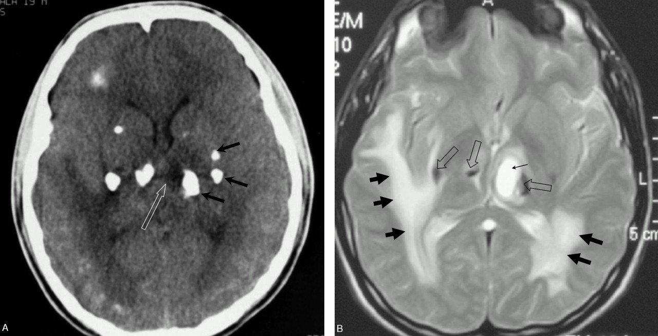
60 yrs old Male asked about Calcified focus in brain 1 doctor answered this and 771 people found it useful. These lesions may be detected in association with the occurrence of seizures or incidentally. Parenchymal Calcifications Parenchymal brain calcifications are often the only imaging finding in NCC.
Calcific Focus In Brain. Connect with a US. Calcific focus in left region of brain.
The most common sites include. Calcific foci in brain Download Here Free HealthCareMagic App to Ask a Doctor All the information content and live chat provided on the site is intended to be for informational purposes only and not a substitute for professional or medical advice. Calcifications are typically 210 mm in diameter well defined and solid Fig.
Dr Henry Knipe and Assoc Prof Frank Gaillard et al. Basal ganglia calcification is a very rare condition that happens when calcium builds up in your brain usually in the basal ganglia the part of your brain that helps control movement. Normal age-related intracranial calcifications.
Brain calcification left untreated. During a routine ultrasound test of my father calcified foci and granulomas were seen in the spleenWhat are the causes of it. Calcific foci in brain.
This inflammation may be due to some infection vascular problem injury or any other trigger. Multiple lacunar ischaemic foci in brain. Board-certified doctor by text or video anytime anywhere.
Is it normal or should he undergo any other tests. Multiple calcifications are formed. Having calcium deposits in my own brain doesnt make me qualified to give advice.
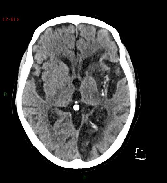
50 off with 15month membership. Intracranial calcifications are common in certain locations and often are of no clinical concern. Brain stones typically manifest as seizures but can also be detected incidentally during brain tomography.
Registered users can ask questions leave comments and earn points for submitting new answers. What is mean of calcific foci. Im no expert on brain calcification.
After being diagnosed with the calcium deposits I havent changed a thing. In the form of focal or diffuse deposits calcification of the brain is detected with MRI in patients with tumors - teratoma meningioma craniopharyngioma intraventricular epindimoma adenoma of the pineal gland. How to dilute calcification from brain.
He is 54 years old. Calcific Focus In Brain. Such lesions likely represent fibrotic reactions to prior infection that have calcified.
Although intracranial calcifications are observed frequently brain stones are less frequently encountered. Calcified Granulomatous Lesions of Brain Whenever an inflammation occurs the tissue affected respond producing an inflammatory exudate and a granuloma is formed. Get your query answered 247 only on Practo Consult.
Testimony of Patient suffered from Multiple Calcific Foci in Brain successfully Treated after our Homoeopathic Medicine TreatmentDrAnkit Srivastava Social. Calcification over 1 cm in diameter or under nine years old may be suggestive. Importantly the calcified lesions may be enriched in vascular tissue.
The two most commonly encountered types of calcification include. 247 visits - just 39. Talk to a doctor now.
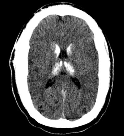
27 yo m. Seen in 23 of the adult population and increases with age. Normal intracranial calcifications can be defined as all age-related physiologic and neurodegenerative calcifications that are unaccompanied by any evidence of disease and have no demonstrable pathological cause.
Brain Pill is one of the only nootropic supplement brand names that has actually experienced the difficulty of running a professional study on its formula. Get the Free App for Members. What is foci in the brain.
Mural and eccentric intravascular calcification. Brain stones or cerebral calculi are large solitary or multiple intracranial calcifications Tiberin and Beller 1963.
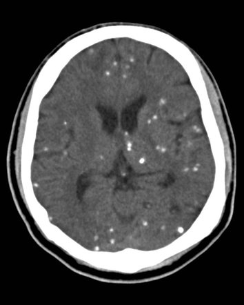
Multiple Intracranial Calcifications Radiology Reference Article Radiopaedia Org
Rail Road Tract Calcification Seen In Sturge Weber Syndrome An Autosomal Dominant Neurocutaneous Syndrome Characteri Port Wine Stain Port Wine Wine Stains
Calcified Neurocysticercosis Lesions Trigger Symptomatic Inflammation During Antiparasitic Therapy American Journal Of Neuroradiology


Intracranial Tumours With Calcification Radiology Reference Article Radiopaedia Org

Dystrophic Calcification In A 73 Year Old Female With A Prior History Download Scientific Diagram
Multiple Intracranial Calcifications Radiology Reference Article Radiopaedia Org

Multiple Intracranial Calcifications Radiology Reference Article Radiopaedia Org

Patient 2 Axial Computed Tomographic Brain Scans Calcified Changes Download Scientific Diagram

Adamantinomatous Craniopharyngioma Ct Brain Demonstrates A Large Suprasellar Predominantly Cystic Mass With Peripheral Ca Pediatric Radiology Pet Ct Radiology
Cortical Calcifications In Ischemic Stroke Radiology Case Radiopaedia Org

Mri Of The Brain Showing Foci Of Calcification Arrows In Bilateral Download Scientific Diagram

Pin By Dr Abuaiad On Brain Head And Neck Brain Tumor Cns Radiology

Falx Cerebri Calcification Radiology Case Radiopaedia Org Radiology Brain Images Sonography
Calcified Neurocysticercosis Lesions Trigger Symptomatic Inflammation During Antiparasitic Therapy American Journal Of Neuroradiology


Multiple Intracranial Calcifications Radiology Reference Article Radiopaedia Org

Cerebral Cortical Calcification Radiology Reference Article Radiopaedia Org
Leukoencephalopathy Cerebral Calcifications And Cysts American Journal Of Neuroradiology
Unilateral Subcortical Calcification A Manifestation Of Dural Arteriovenous Fistula American Journal Of Neuroradiology


Neuroradiology On The Net Congenital Cytomegalovirus Infection Radiology Imaging Radiology Neurology
0 Response to "Calcific Foci In Brain"
Post a Comment