
Ischemic Foci In Brain
Microvascular ischemic disease is a term thats used to describe changes to the small blood vessels in the brain. There is no acute ischemic infarction and the major intracranial arterial flow voids are patent.
Acute And Chronic Brain Infarcts On Mr Imaging In A 20 Year Old Woman With Acute Posterior Multifocal Placoid Pigment Epitheliopathy American Journal Of Neuroradiology
If the patient is given and infusion of gadolinium the spots will appear even brighter on the MRI film.

Ischemic foci in brain. Individuals with white matter foci may or may not. Has your father ever suffered from heart attack. Ischemia means reduced blood flo.
Drug overdose if it leads to low blood pressure with subsequent hypoxic-ischemic brain injury could theoretically lead to multiple spots in the brain with a specific pattern called a watershed pattern. They usually indicate physiological changes caused by disease processes infections or the normal aging process. 50 year old female presented complaining of headaches 3-4 times per week.
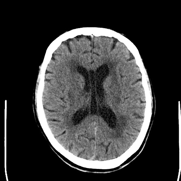
It can be either caused by thrombosis or embolism. These are signs of microvascular ischemic disease in the brain tissue. From one-third to 80 percent of MRI scans performed on patients older than 65 show T2 hyperintense foci as of 2015.
White matter foci commonly appear on a magnetic resonance imaging as bright white spots on the part of the brain that contains nerve cells covered with lipid tissue known as myelin. These four- 2 in cerebral white matter and another two in peri-ventricular region -ischemic lesions can be due to emboli arising either from arteries from the neck or heart. Foci such as these can also occur in the setting of migraine headaches.
2 Scattered foci of increased signal in white matter most likely ischemic in nature. Foci on an MRI are periventricular white matter lesions evidence of changes in a patients brain that appear on the MRI as white spots states Timothy C. Other investigations are sometimes necessary to distinguish the.
Headaches are present upon waking in the morning and occasionally late in the day. Multiple foci of TFLAIR hyper intensity involving the subcortical white matter are unchanged. In case you arent familiar with the terms used here.
Hyperintense means there is a spot of increased signal on the imaging. Focal brain ischemia occurs when a blood clot has occluded a cerebral vessel. There are a few punctate periventricular white matter lesions that are also stable.
Microvascular ischemic brain disease describes conditions that affect the small blood vessels in the brain. Two common sources of embolization to the brain are the heart and internal carotid artery. Brain pHi and cortical blood flow of the ischemic penumbra measured 661 - 006 and 319 - 92 mL100 g per minute.
Citation needed Global brain ischemia. These conditions include stroke cerebral. Chronic ischaemic foci is a term thats largely used in relation to pathologies in the brain.
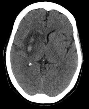
The corpus callosum is normal in thickness and signal intensity. Your info suggests brain vascular problems that are causing ischemia reduced blood flow to brain tissue which until now has been mild or transient b. A hyperintense focus in the right frontal lobe in the white matter is a common finding seen on MRI.
Focal brain ischemia lasting more than 3060 min in the majority of cases produces a cerebral infarct. The foci typically appear in areas with higher levels of fluid. Family has history of severe migraine headaches mother and brother.
Although the number of foci has been reported to correlate with age and several risk factors the degree of observer variability in qu. Foci of high signal in the cerebral white. Read More 1 doctor agrees.
Focal brain ischemia reduces blood flow to a specific brain region increasing the risk of cell death to that particular area. Changes to these vessels can damage white matter. Common neuropathological characteristics of ischemic.
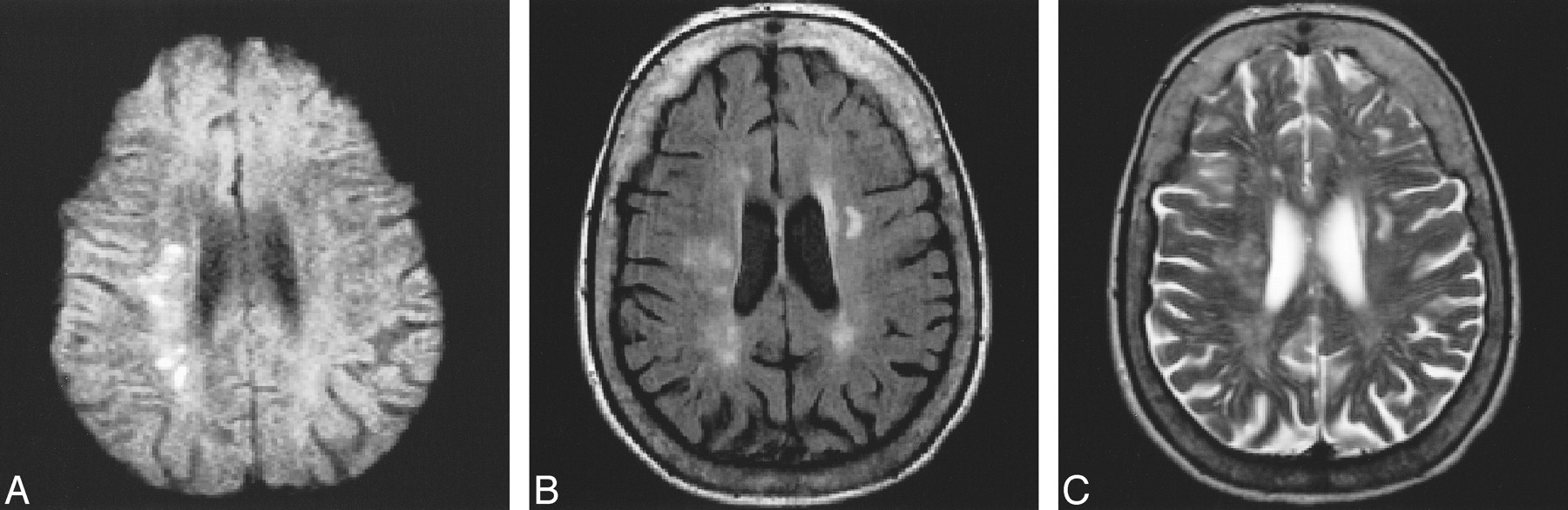
In this MRI of the brain there are multiple white spots that appear very brightly in the brain tissue. Patient has experienced migraine. The term chronic suggests that the problem is one of long duration.
This needs to be correlated with the patients clinical symptoms. Over 3 hours there was normalization of pHi in the majority of the penumbra due to increases in cortical blood flow. Within the ischemic penumbra acidic foci developed with an.
The different causes of white matter hyperintensities can be determined to some extent by interpretation of various sequences of the MRI. Foci of high signal in the cerebral white matter are common incidental findings on MR images of the brain of control subjects or patients with a variety of diseases. Brain infracts may be a consequence of either embolic or thrombotic processes.
A thrombotic arterial brain infarct may result from severe atherosclerosis of an artery leading to vascular occlusion and a severe drop in local cerebral blood flow.

A Mri T2flair Of The Brain On Admission Showing Bilateral Occipital Download Scientific Diagram

Cerebral Infarction An Overview Sciencedirect Topics
Blood Brain Barrier Reperfusion Injury And Hemorrhagic Transformation In Acute Ischemic Stroke Neurology

The Radiology Assistant Imaging In Acute Stroke
How Covid 19 Effects The Brain In Neuroimaging Daic
Stroke Imaging Practice Essentials Computed Tomography Magnetic Resonance Imaging

Brain Mri Flair Scans With Subacute Ischemic Lesions In Left Frontal Download Scientific Diagram

White Matter Lesion An Overview Sciencedirect Topics
Https Pubs Rsna Org Doi Pdf 10 1148 Rg 2016160031
Diffusion Weighted Imaging Of Patients With Subacute Cerebral Ischemia Comparison With Conventional And Contrast Enhanced Mr Imaging American Journal Of Neuroradiology
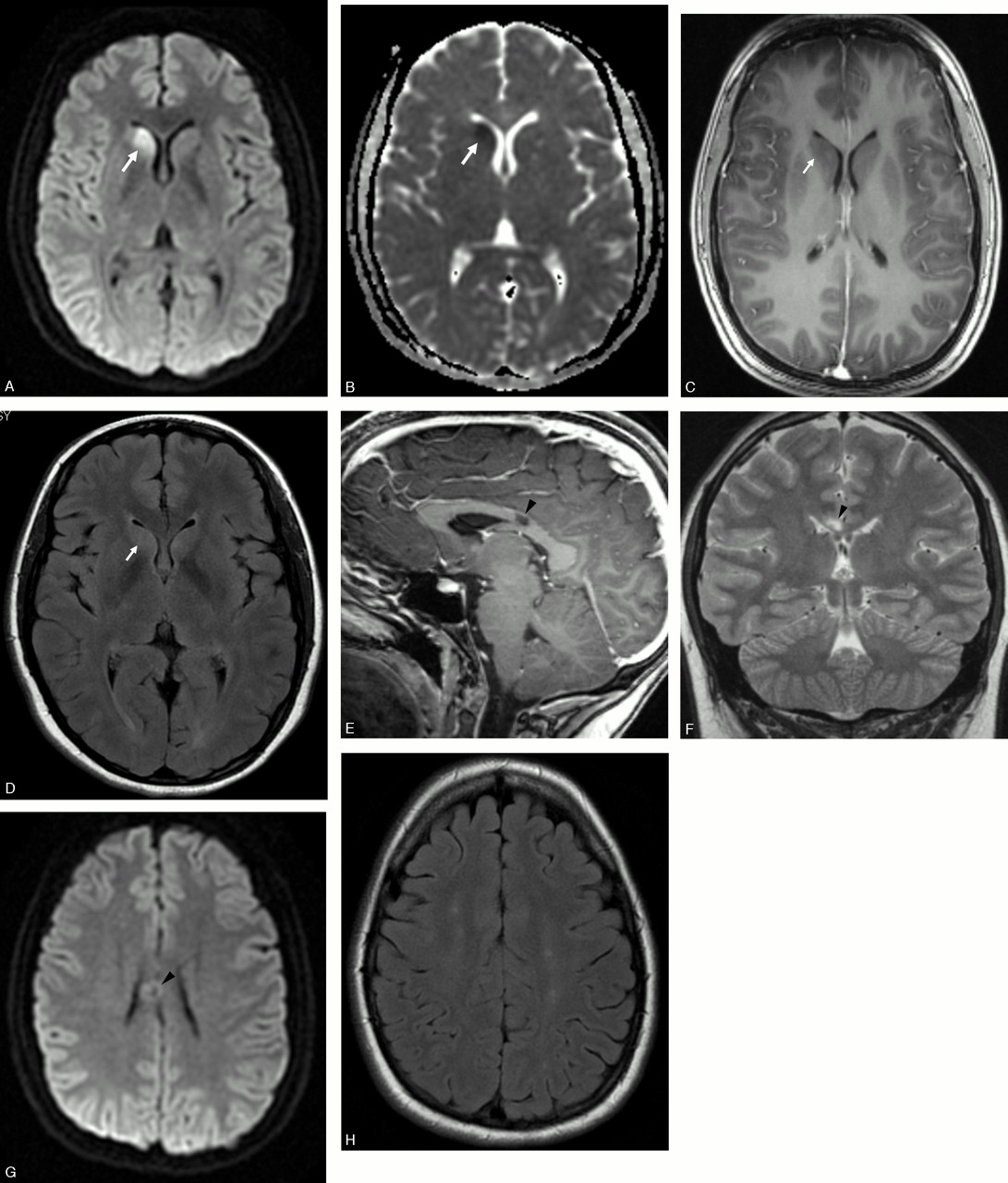
Reversible And Benign Lesions Of Splenium Of The Corpus Collosum Practical Neurology

Diffusion Weighted Imaging Of Patients With Subacute Cerebral Ischemia Comparison With Conventional And Contrast Enhanced Mr Imaging American Journal Of Neuroradiology


White Matter Hyperintensities On Mri Artefact Or Something Sinister

Mri Of Ischemic Lesions Abnormal Signal Changes In Multiple Brain Download Scientific Diagram

Prevention Of Stroke In Patients With Silent Cerebrovascular Disease A Scientific Statement For Healthcare Professionals From The American Heart Association American Stroke Association Stroke

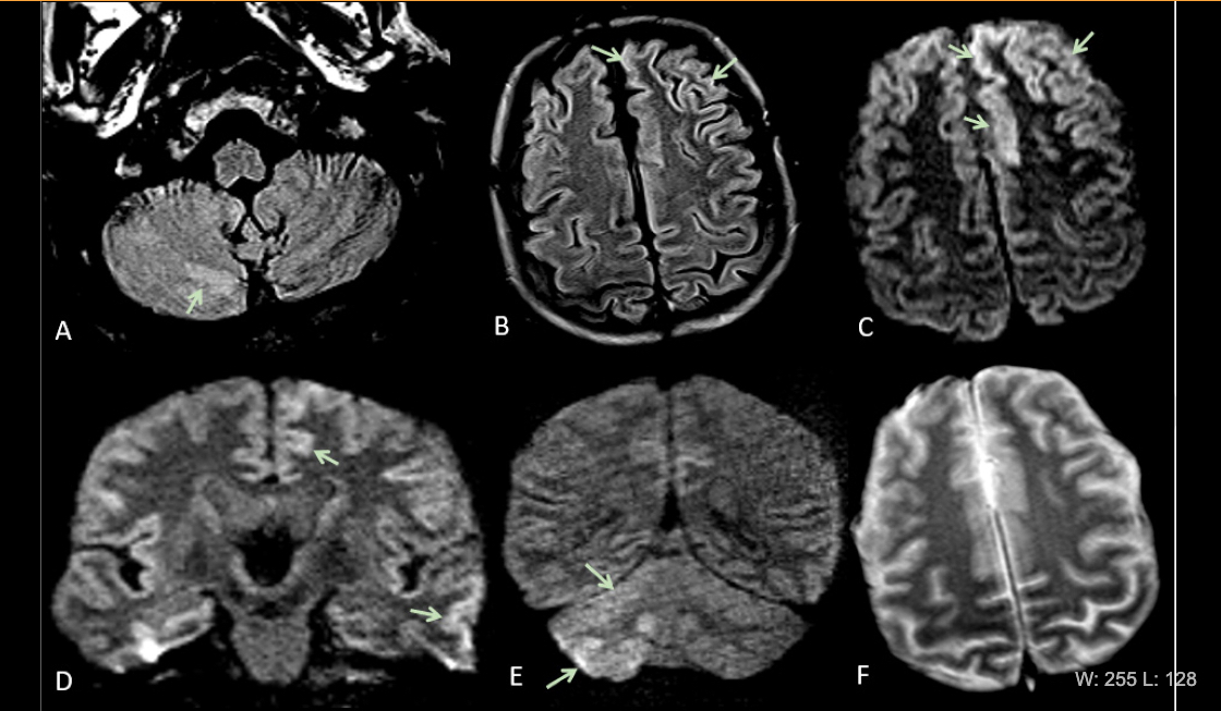
Association Between Remote Diffusion Weighted Imaging Lesions And Cerebral Small Vessel Disease In Primary Intracerebral Hemorrhage Xu 2019 European Journal Of Neurology Wiley Online Library

Asymptomatic Acute Ischemic Lesions In Intracerebral Hemorrhage Its Frequency Mri Features And Risk Factors Journal Of The Neurological Sciences
Fig 1 Brain Mri Findings In Neurologically Asymptomatic Patients With Infective Endocarditis American Journal Of Neuroradiology

Chronic Small Vessel Ischemia Radiology Case Radiopaedia Org
0 Response to "Ischemic Foci In Brain"
Post a Comment